![]()
![]()
![]()
|
|
|
|
The eye is like a camera. When you look at an object, light rays are reflected from the object to the cornea. The light rays are bent, refracted and focused by the cornea, lens, and vitreous. The lens helps bring rays of light to come to a sharp focus on the retina. The resulting image on the retina is upside-down. Here at the retina, the light rays are converted to electrical impulses which are then transmitted through the optic nerve, to the brain, where the image is translated and perceived in an upright position.
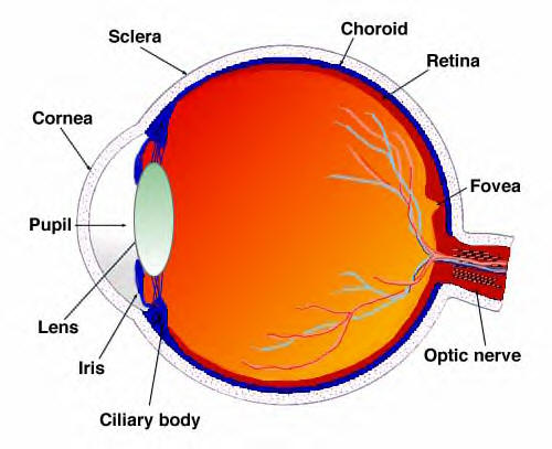
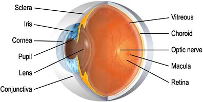
cross-section of the eye
The external layer Cornea - the transparent front "window" of the eye that covers the iris and pupil and provides most of the eye's optical power. Light must pass through the cornea when it enters the eye. Attached to the sclera are the extraocular muscles that move the eye. Sclera - white of the eye which forms part of the supporting wall of the eyeball. The sclera is continuous with the cornea. The intermediate layer Choroid - contains the blood vessels that supply blood to structures of the eye - the front part of the choroid contains two structures: Ciliary body - a muscular area attached to the lens. It contracts and relaxes to control the size of the lens for focusing. Iris - the coloured part of the eye. The colour of the iris is determined by the colour of the connective tissue and pigment cells. Less pigment makes the eyes blue, more pigment makes the eyes brown. This circular muscle controls the size of the pupil so that more or less light, depending on conditions, is allowed to enter the eye. The internal layer Retina - the sensory part of the eye. The lining of the rear two-thirds of the eye, the retina converts images from the eye's optical system into electrical impulses sent along the Optic Nerve to the brain. The macula is the highly sensitive area of the retina. The macula is the part of the retina most used and is responsible for acute central vision. We use our macula to read or to stare intently at an object.
Three fluid chambers Anterior chamber - between cornea and iris Posterior chamber - between iris, zonule fibres and lens Vitreous chamber - between the lens and the retina
All these parts must be functioning correctly for the eye to transmit impulses along the optic nerve so that the brain can assemble a clear picture. If not, vision is impaired.
/ Contact / Our People / Eye Conditions / Links / Cataract Surgery / Eye Surgery / Glaucoma / Retinal Detachment / Refractive Surgery / Clear Lens Surgery / Macular Degeneration / News
Page last updated on Monday, 29 August 2005 18:54:41 |
|
Highgate Ophthalmic Practice
|